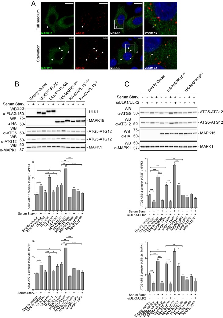
Figure 7.
MAPK15 controls early phases of autophagosomal biogenesis through ULK1/2. A, HeLa cells were transfected with HA-MAPK15WT plasmid. After 48 h, cells were incubated for 4 h in full medium or serum starvation medium. MAPK15 (green) and ATG12 (red) were labeled to monitor their subcellular localization. Nuclei were visualized in blue. Colocalization is shown in yellow and indicated by white arrowheads. Reference bars correspond to 10 μm. Representative images from three different experiments are shown (n = 3). B, HeLa cells were transfected with plasmid encoding for ULK1WT-FLAG, ULK1KD-FLAG, HA-MAPK15WT, HA-MAPK15AXXA, HA-MAPK15KD, or the empty vector. After 24 h, cells were incubated for 4 h in full medium or serum starvation medium. Total lysates were analyzed by Western blot (WB) analysis for endogenous ATG5 and ATG12 (n = 3). ATG12 levels were plotted on the graph (lower panel). ATG5 levels were plotted on the graph (middle panel). C, HeLa cells were transfected with ULK1 plus ULK2 siRNA or SCR siRNA and, after 48 h, further transfected with plasmid encoding for HA-MAPK15WT, HA-MAPK15KD, or the empty vector. After 24 h, cells were incubated for 4 h in full medium or serum starvation medium. Total lysates were analyzed by Western blot analysis for endogenous ATG5 and ATG12 (n = 3). ATG12 levels were plotted on the graph (lower panel). ATG5 levels were plotted on the graph (middle panel).

