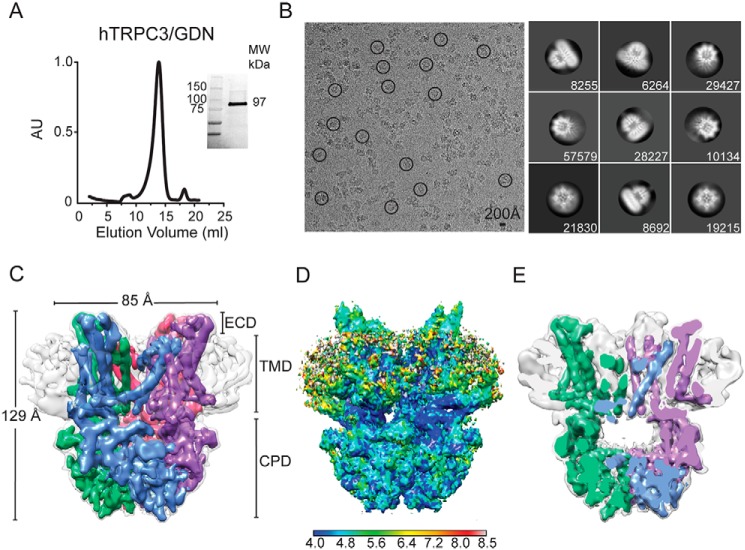
Figure 1.
Cryo-EM structure of full-length human TRPC3 at 5.8 Å. A, size-exclusion chromatography profile of digitonin-solubilized and GDN-purified TRPC3 from Sf9 cells. Inset, stain-free protein on SDS-PAGE gel corresponds to TRPC3 monomer (97 kDa). B, left, micrograph after motion correction of TRPC3 in GDN micelles (TRPC3GDN), taken on an FEI Polara microscope. Note that particles are monodisperse and some are circled in black. Right, representative 2D class averages of TRPC3GDN. Particles were aligned and classified in RELION 2.1. The number of particles in each class is shown in the lower right corner of each box. C, electron density map of TRPC3GDN tetrameric assembly. GDN micelle is denoted in light gray at a higher threshold than the four subunits, colored in blue, green, pink, and purple. D, side view of TRPC3GDN with local resolution calculated in ResMap indicated by the heat map scale bar. High- to low-resolution runs as blue to red, from 4.0 to 8.0 Å. E, side view cross-section of the tetrameric TRPC3GDN highlighting the hollow inner chamber below the transmembrane domain. AU, arbitrary units; ECD, extracellular domain.

