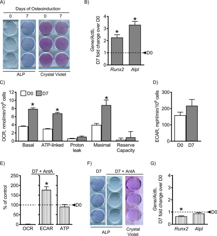
Figure 1.
Mitochondrial OxPhos is necessary for osteogenesis. C3H cells were osteoinduced for 0 and 7 days using medium containing ascorbate and β-glycerol phosphate. Staining for osteoblast marker ALP and total cell crystal violet stain showed that cells up-regulated ALP at day 7 (D7), indicating osteogenesis (A). Gene expression of osteogenic markers Runx2 and Alpl is significantly increased at day 7, further indicating osteogenesis (B). OCR, a readout of mitochondrial OxPhos, was significantly increased in basal, ATP-linked, and maximal respiration at day 7 (C), whereas there was no change in glycolytic rates as measured by ECAR (D). When treated with AntA, a mitochondrial OxPhos inhibitor, at 0.5 μm, mitochondrial OxPhos is undetectable, and glycolysis is significantly increased, indicating a glycolytic shift with AntA, without noticeable effect on ATP levels (E). Treatment with AntA inhibited osteogenesis as shown by a reduced ALP stain and reduced gene expression of osteogenic markers (F and G). This indicates that mitochondrial OxPhos is required for osteogenesis. Dashed lines indicate day 0 (D0). Data are means. Error bars represent S.E. (n = 3–5). *, p < 0.05 compared with day 0 control.

