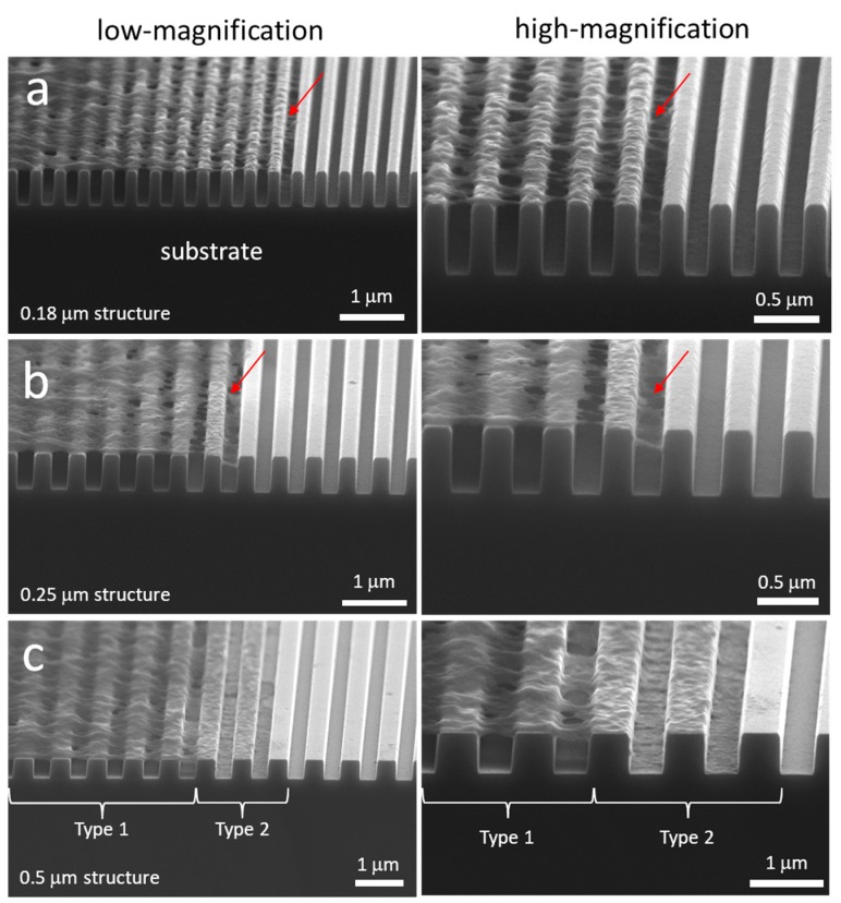
Figure 7.
SEM micrographs 70° tilted of cross-sectioned cells on comb structures with line widths of (a) 0.18 μm, (b) 0.25 μm, and (c) 0.5 μm. Two distinct types of cell adhesion morphologies are observed: Type 1—Adherent cells on 0.18 μm and 0.25 μm structures only contacted the top portion of lines but did not fill the trench gaps; and Type 2—Cells exhibit conformal surface coverages. Both morphology types were observed for adherent cells on the 0.5 μm comb structures. All cells were incubated on the structures for 24 h. Cell concentration was ~5 × 10⁵ cells/mL.

