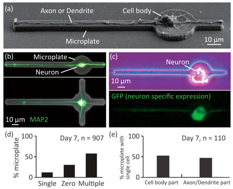
Figure 3.
Culturing single neurons on the microplates. (a) A SEM image of a single neuron on a microplate; (b) Immunostained neurons on microplates; green: MAP2; (c) GFP expression under control of excitatory-neuron-specific CaMKII promoter; (d) Histogram of number of cells on individual microplates; (e) Histogram of positions of cell body of single neurons on microplates.

