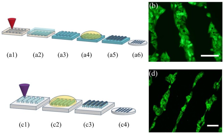
Figure 8.
Diagram of PMMA laser etching for the fabrication of µCP master molds: (a1) Laser etching of PMMA to obtain a mastermold, (a2) PDMS is poured onto the PMMA structured master, (a3) the stamp is peeled off the master, (a4) the stamp is incubated with an ink solution, (a5) the excess of solution is removed, (a6) the inked stamp is placed onto a glass coverslip and the protein pattern is suitable for cell culture. (b) HepG2 cells grow selectively onto the collagen I protein pattern (green is calcein (live cells), scale bar: 100 µm. Diagram of Loctite DLW for the fabrication of µCP stamps in one single step: (c1) Selective laser crosslinking of Loctite to obtain the desired stamp, (c2) the stamp is incubated with an ink solution, (c3) the excess of the solution is removed, (c4) the inked stamp is placed onto a glass coverslip and the protein pattern is suitable for cell culture. (d) HepG2 cells onto the transferred protein pattern (green is phalloidin as actin cytoskeleton marker), scale bar: 100 µm.

