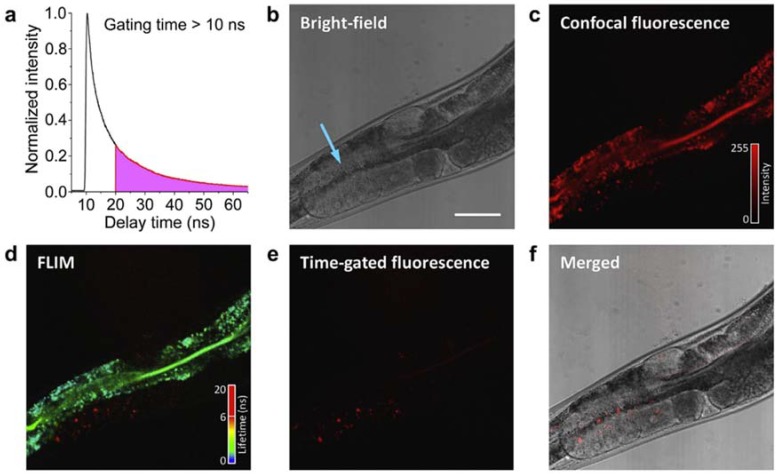
Figure 2.
Observation of green fluorescent protein (GFP):Yolk lipoprotein complexes (YLC)-fluorescent nanodiamonds (FNDs) in Caenorhabditis elegans by fluorescence lifetime imaging microscopy (FLIM). (a) A fluorescence decay time trace of 100-nm FNDs excited by a picosecond pulsed laser; (b) bright field image, blue arrow indicates the site of injection; (c) confocal fluorescence image; (d) FLIM image; (e) time-gated fluorescence image at a delay greater than 10 ns; and (f) merged image of (b,e). Scale bar: 50 µm. Reprinted with permission [65]. Copyright (2012) Elsevier Ltd.

