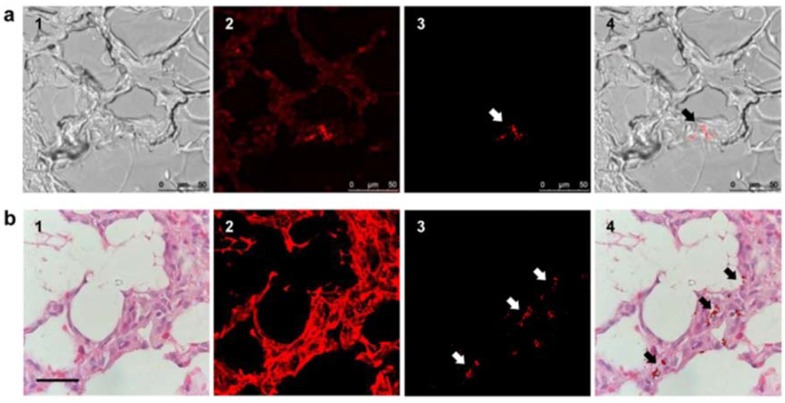
Figure 3.
Fluorescence imaging of FND-labeled mesenchymal stem cells (MSC) in pig tissues. (a) Fast screening of a lung tissue before deparaffinization to find FND-labeled MSCs: (1) bright-field image, (2) fluorescence image without time-gating, (3) fluorescence image with time-gating at delays greater than eight nanoseconds, and (4) merged images of (1,3). Scale bar: 50 µm. (b) Identification of FND-labeled MSCs in a lung tissue section by confocal microscopy: (1) Hematoxylin & Eosin staining image, (2) confocal fluorescence image without time gating, (3) time-gated confocal fluorescence image, and (4) merged image of (1,3). Scale bar: 20 µm. Reprinted with permission [63]. Copyright (2017) Springer Nature.

