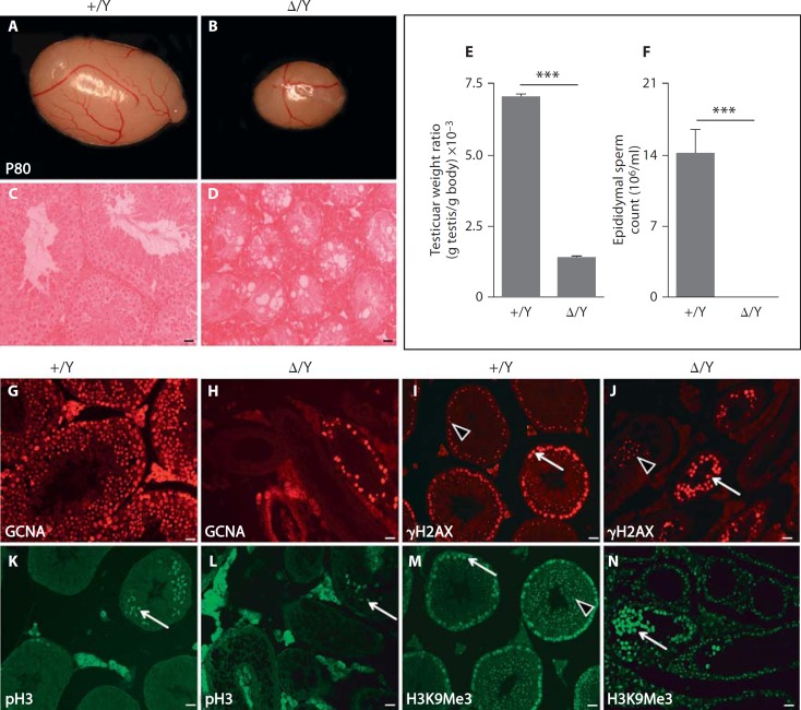
Fig. 2.
Reduction in testis size and absence of spermatozoa in GilzΔ/y mutant mice. P80 adult testes from GilzΔ/y (Δ/Y, n = 11) (B) mice showed an 80% reduction in weight (E) compared to control (+/Y, n = 6) littermates (A). H&E staining of testes sections (C, D) revealed complete absence of mature spermatozoa and elongated spermatids in Δ/Y testes. F Sperm count analysis revealed a complete absence of epididymal sperm in Δ/Y mutant animals. G, H Immunostaining using GCNA antibody revealed a drastic reduction in the number of germ cells present in mutant seminiferous tubules (H) compared to +/Y tubules (G). I, J Anti-γH2AX staining revealed the reduced presence of early meiotic spermatocytes (whole nucleus staining, arrows) and few sparse pachytene spermatocytes (punctate staining, arrowheads) within Δ/Y testis (J) compared to wild type (I). Few metaphasic cells are also present in Gilz mutants as shown by histone H3Ser10 phosphorylated (pH3) positive cells (compare staining in K and L). M, N Sex chromatin of round spermatids is detected by punctate H3K9Me3 staining in control tubules (M, arrowhead) but is absent in mutant tubules (N); arrows show staining of whole nucleus of spermatocytes. For E and F, a minimum of 6 animals were analyzed for each genotype. Results are mean ± SEM, *** p < 0.0001 versus controls. Scale bars = 20 µm.

