
Figure 4.
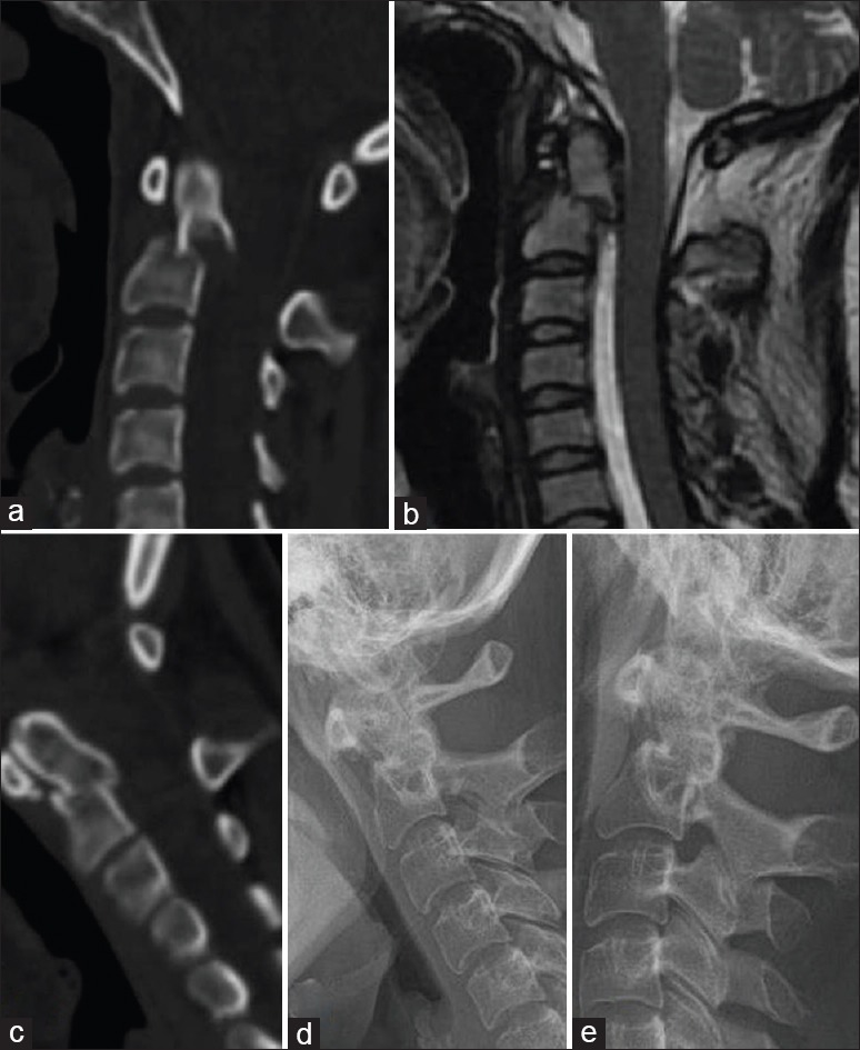
A 24-year-old female patient with Type II odontoid fracture. (a) Type II odontoid fracture >6 mm in the sagittal sequence of computerized tomography is shown. (b) View of T2 sagittal magnetic resonance imaging. (c) Moderate reduction was seen after halo in sagittal computerized tomography. (d and e) No sign for instability was reported in the dynamic graphics end of the 1st year