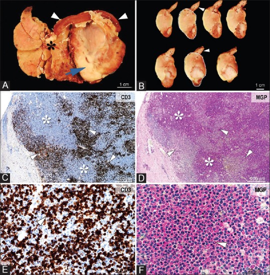
Figure 4.

Gross and microscopic images of lymphoma and a plasma cell tumor involving the liver. Grossly, the tumor (Panel A, arrow) infiltrates and displaces the liver (Panel A, asterisk) and spleen (Panel A, arrowhead). On cross-section, the tumor (Panel B, demarked area) is heterogeneous, white-beige, friable, with large areas of necrosis and hemorrhage (Panel B, arrowheads). Histologically, the tumor consists of large areas of neoplastic plasma cells (Panel C and D, asterisks), associated with coalescing foci of CD3 immunopositive lymphocytes (Panel C-E, arrowheads). The densely packed plasmacytoid cells are round to oval, with variably sized, and eccentrically placed nuclei with coarse chromatin, a prominent Golgi area, and cytoplasm that stain intensely for methyl green-pyronin (Panel F, arrowhead). CD3 immunolabeling, objective ×4 (Panel C) and × 40 (Panel E). Methyl green-pyronin stain for plasma cells, objective ×4 (Panel D) and ×40 (Panel F)
