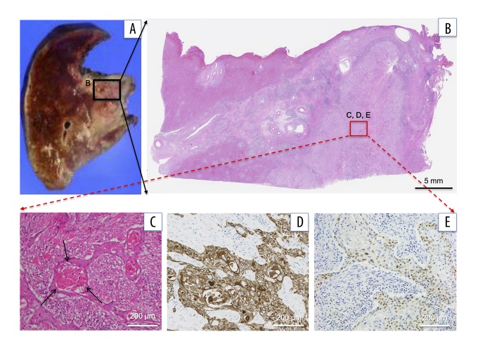
Figure 3.
Immunohistopathological findings of primary liver tumor specimens. (A) The resected specimens of the liver: (B) low power field and (C) high power field. Microscopic findings of resected tumor. Most (>99%) of this liver tumor is composed of well-differentiated squamous cells. The tumor is composed of squamous cells with keratinization that forms cancer pearls (H&E staining, arrows). (D) Squamous cells show diffusely positive CK5/6 staining immunohistochemically. (E) Squamous cells show positive p40 staining immunohistochemically. H&E – hematoxylin and eosin.

