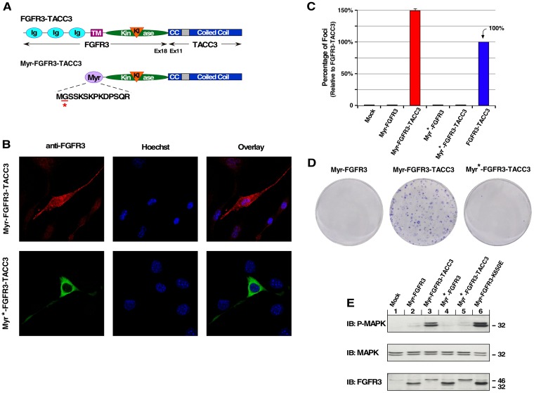
Figure 2. Plasma membrane-localized FGFR3-TACC3 results in cell transformation.
(A) Schematic of FGFR3-TACC3 and Myr-FGFR3-TACC3 fusion proteins. For the membrane-localized fusion construct, the extracellular and TM domains of FGFR3 are replaced with a myristylation sequence (Myr) derived from c-Src (Myr-FGFR3-TACC3). Mutation of underlined residue to Ala (A) results in cytoplasmic-localized FGFR3-TACC3 (Myr*-FGFR3-TACC3). (B) Representative confocal micrographs of NIH3T3 cells stably expressing the indicated constructs, using FGFR3 immunostaining (P-18). Secondary antibodies were either donkey anti-goat AlexFluor488 or donkey anti-goat AlexaFluor594. Nucleus is visualized with Hoechst 33342. (C) Transformation of NIH3T3 cells by FGFR3 and FGFR3-TACC3 derivatives. Number of foci were scored, normalized by transfection efficiency, and quantitated relative to FGFR3-TACC3 ± SEM. Assays were performed a minimum of three times per DNA construct. (D) Representative plates from a focus assay are shown, with transfected constructs indicated. (E) HEK293T cell lysates expressing FGFR3 or FGFR3-TACC3 derivatives were immunoblotted for phospho-MAPK (T202/Y204; top), MAPK (second panel), and FGFR3 (bottom).

