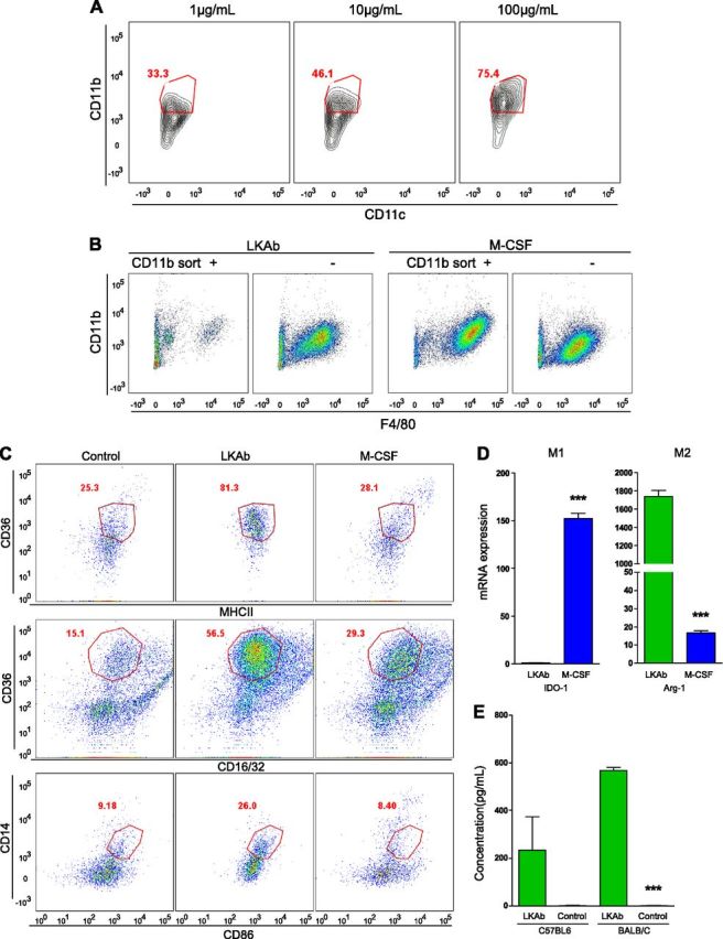
Figure 3.

LKAb induced anti-inflammatory M2 macrophage differentiation. A) Mouse bone marrow cells were incubated with LKAb at the indicated concentrations (1–100 µg/ml) for 6 d. The cells were then stained with anti-CD11b and -CD11c and analyzed by FACS. Red outline: positive macrophage populations. LKAb induced differentiation of CD11b+ macrophages, but not CD11c+ dendritic cells, in a dose-dependent manner. B) Mouse total bone marrow cells were separated by using CD11b-specific magnetic beads, and the isolated CD11b+ or CD11b− populations were incubated with LKAb or M-CSF for 6 d. The cells were then stained with anti-CD11b and -F4/80 and analyzed by FACS. Macrophages differentiated mainly from the CD11b− population. C) Mouse bone marrow was incubated with medium, LKAb, or M-CSF for 6 d. Cells were then stained with anti-CD16/32 and -CD86 as M1 macrophage markers, and anti-CD36, -MHCΙΙ, and -CD14 as M2 macrophage markers. Red outline: M2-type-specific populations. The macrophages induced by LKAb selectively expressed M2 type markers. D) Mouse bone marrow was induced by LKAb antibody or M-CSF for 6 d. Cells were harvested, and total RNA was extracted for qRT-PCR analysis. IDO1 was used as an M1-specific gene and ARG-1 as an M2-specific gene. qRT-PCR showed high ARG-1 mRNA expression in macrophages induced by LKAb but only low levels of IDO1. E) Macrophages induced by the LKAb in vivo expressed M2 cytokines. LKAb and PBS were injected intraperitoneally into C57BL/6 or BALB/c mice 3 times/wk for 2 wk, and IL-10 levels in the serum were measured. Mice treated with LKAb showed dramatically increased levels of IL-10, one of the major anti-inflammatory cytokines. ***P < 0.0005; Student’s t test.
