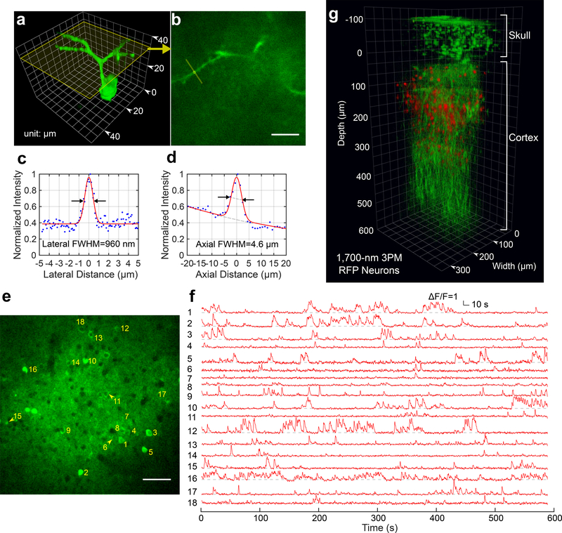
Figure 2|. Through-skull Imaging of Neural Structure and Function.
a, 3D reconstruction of a GCaMP6s-labeled neuron located about 140 μm below the cortical surface of a transgenic mouse (CamKII-tTA/tetO-GCaMP6s, 10 weeks; similar measurements were performed on 2 mice with 3 neurons each, in total n=6) imaged by 1320-nm 3PM through the intact skull (thickenss ~ 100 μm). Apical dendrites can be clearly observed for resolution estimation.
b, A cross section of the 3D stack in (a), with its location indicated by the yellow frame in (a). The locations for lateral and axial resolution measurement on the apical dendrite are indicated by the yellow line and circle, respectively (similar results n=6). Scale bar, 10 μm.
c, Lateral intensity profile measured along the yellow line in (b), fitted by a Gaussian profile for the estimation of the lateral resolution (similar results n=6).
d, Axial intensity profile measured in the region within the yellow circle in (b), fitted by the sum of two Gaussian profiles, with one broad, off-centered Gaussian profile to corrected for the uneven baseline, and the other for the central peak (similar results n=6).
e, High resolution image of a site for through-skull activity recording in an awake, GCaMP6s-labeled transgenic mouse (CamKII-tTA/tetO-GCaMP6s, female, 8 weeks, similar results n=5). The recording site is about 275 μm below the cortical surface and the FOV was 320 μm × 320 μm (256×256 pixels per frame). Scale bar, 50 μm
f, Spontaneous activity traces recorded under awake condition from the indexed neurons in (e), acquired at 8.49 Hz frame rate (similar results n=5). The repetition rate used for imaging was 800 kHz, and the average power under the objective lens was 44 mW. Each trace is normalized to its baseline and low-pass filtered by a hamming window of 1.06 s time constant. The same site was visited 8 times over a period of 4 weeks after the skull preparation, with cumulatively over 6 hours of recording time (data from other imaging sessions are shown in Supplementary Fig. 5).
g, 3D reconstruction of through-skull imaging of a cortical column of red fluorescent protein (RFP) labeled neurons in a Brainbow mouse (B6.Cg-Tg(Thy1-Brainbow1.0)HLich/J, male, 12 weeks, similar results n=2). The red channel is 3PE fluorescent signal from RFP and the green channel is THG. The zero depth is set just beneath the skull.

