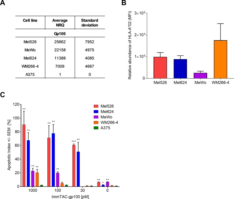
Fig 3. ImmTAC-mediated killing capacity is dependent on the levels of target antigen presentation.
(A) gp100 mRNA levels measured in specified cell lines using qRT-PCR. Expression is presented as Normalised Relative Quantity (NRQ) relative to housekeeping genes (RPL32 and HPRT1). NRQ = (RQ target gene/geometric mean RQ housekeeping genes) x 104, RQ = Efficiency-CT. n = 3, 4, 5, 3 and 2 for Mel526, Mel624, MeWo, WM266-4 and A375, respectively. (B) Levels of HLA-A*02 protein on the cell surface of specified cell lines measured by flow cytometry (mean fluorescence intensity (MFI) adjusted to the isotype control). n = 8, 9, 4 and 3 for Mel526, Mel624, MeWo, VM266-4 and A375, respectively. (C) Killing capacity of ImmTAC-gp100-redirected T cells assessed using the IncuCyte assay. A range of cell lines expressing different levels of gp100-HLA-A*02 complexes were incubated with decreasing concentrations of ImmTAC-gp100. The apoptotic index was determined by calculating the % ratio of apoptotic cells to the total number of target melanoma cells at the experimental endpoint (52 hours). n = 2–3. Statistical differences in apoptotic index between Ag+ (Mel 526, Mel624, MeWo and WM266-4) and Ag- (A375) cell lines were individually measured at each concentration of ImmTAC molecule using an unpaired T-test where *** p<0.0001, **p<0.01 and *p<0.05. If unmarked, results were not significant.

