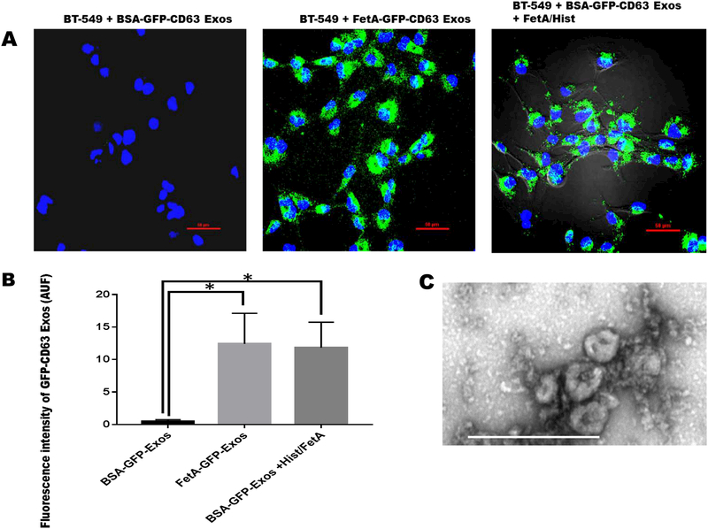
Figure 1. Uptake of GFP-CD63 labeled exosomes by BT-549 breast carcinoma cells.
BT-539 cells were seeded in 8-chambered glass slides (1 × 10⁵ cells/chamber), and incubated for 24 h in complete medium and which the medium was replaced with SFM. Purified exosomes (100 μg/ml) secreted from BT-CD63 cells in the presence of BSA (negative control) or fetuin-A (positive control) in SFM were incubated with the BT-549 cells (2 μg/chamber) to monitor uptake. Exosomes secreted from BT-CD63 in the presence of BSA were also incubated with fetuin-A/histone H2A and re-purified (100 μg/ml) and finally incubated with BT-549 cells (2 μg/chamber) for uptake studies (Fig. 1A; scale bar = 50 μm). Mean arbitrary units of fluorescence ± SD were quantified by NEARS for each of the three measurements (Fig. 1B *P < 0.05; N = 6). Transmission electron micrograph of the purified exosomes obtained as described (14) is represented by Fig. 1C (scale bar is 500 nm).

