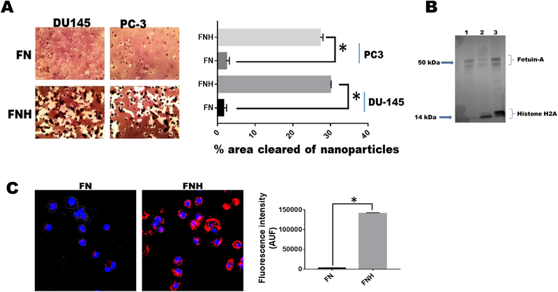
Figure 2. Histones mediate the uptake of hydroxyapatite nanoparticles by tumor cells.
Hydroxyapatite-nanoparticles (2 mg/ml) were incubated with either 2 mg/ml of fetuin-A (FN) or fetuin-A (2 mg/ml) and histones (100 μg/ml) (FNH) in 10 ml of HBSS for 24 h at 4°C. The particles were centrifuged at 700 x g for 5 min to pellet large aggregated particles. The nanoparticles (colloidal) that remained suspended (9 ml) were washed 3X with HBSS, each time pelleted by centrifugation (5,000 x g for 5 min). The final pellets were suspended in 9 ml of complete medium and added to 96-well microtiter plates (100 μl/well) and the tumor cells (1,000 cells/well) added to the lawn of nanoparticles. After 48 h of incubation (37°C in humidified CO2 incubator), the cells were photographed and the % of areas cleared of nanoparticles quantified using NEARS (Panel A; *P < 0.001; N = 6). In panel B, the nanoparticles after the final wash in HBSS, were boiled in Laemmli sample buffer and resolved in NUPAGE and stained with colloidal Coomassie blue (lane 1-FN; lanes 2 and 3-FNH). In panel C, the fetuin-A was labeled with rhodamine isothiocyanate prior to incubation with hydroxyapatite nanoparticles (FN) or the nanoparticles and histones (FNH). The uptake of the nanoparticles (AUF) monitored using A1R confocal microscope and quantified by NEARS (*P < 0.001; N = 6).

