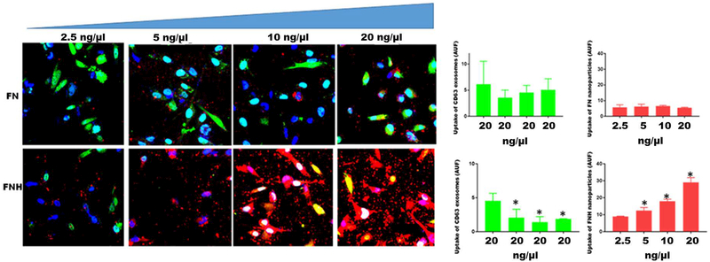
Figure 6. FNH nanoparticles competitively inhibits the uptake of exosomes by breast carcinoma cells.
MDA-MB-231 breast carcinoma cells were plated (2 × 10⁵ cells/chamber) in triplicates in 8-chambered glass slides. After an overnight attachment, complete medium in the chambers was replaced with SFM. GFP-CD63 labeled exosomes were added to each chamber (20 ng/μl) of three separate slides containing cells. To the upper four chambers of each slide, increasing concentrations of FN nanoparticles (2.5–20 ng/μl) in SFM were added. To the lower four chambers, increasing concentrations of FNH nanoparticles (2.5–20 ng/μl) in SFM were added. After 1 h of incubation at 37°C, the chambers were removed and the cells fixed in 4% formalin and a drop of slow-fade with Dapi added and cover-slipped. Uptake of exosomes (green channel) and nanoparticles (red channel) by the cells was monitored by confocal microscopy (Nikon A1R) and arbitrary units of fluorescence quantified using NEARS software as described in Materials and Methods. The bars represent means ± SD (*P < 0.05; N = 3; one way ANOVA)

