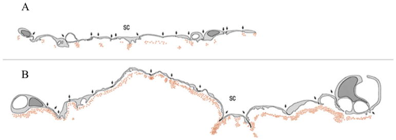
Figure 2.
Schematic of inner wall endothelium of monkey eye perfused with colloidal gold. (A) shows a control eye with punctate distribution of colloidal gold; (B) shows an eye perfused with H-7 where the distribution of colloidal gold is much more uniform81.

