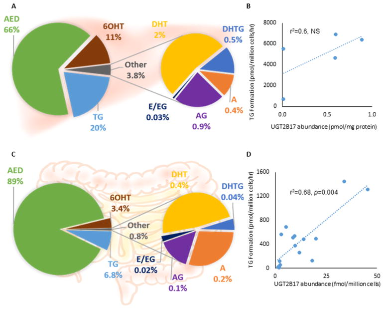
Fig. 4. Testosterone metabolism in primary human hepatocytes and enterocytes.
A&C. Percent of quantified testosterone metabolites in (A) hepatocytes (n=5) and (B) enterocytes (n=16) after incubation in 50 μM testosterone for 60 and 120 minutes, respectively; B&D. UGT2B17 abundance vs. TG formation correlation in (B) hepatocytes (pmol/ mg membrane protein) and (D) enterocytes (fmol/ million cells). UGT2B17 quantification differed due to preexistence of BSA in enterocytes.
TG: testosterone glucuronide, DHT: dihydrotestosterone, DHTG: DHT glucuronide, A: androsterone, AG: androsterone glucuronide, AED: androstenedione, 6OHT: 6-hydroxy testosterone, E: eticholanolone, EG: eticholanolone glucuronide, NS: non-significant

