Fig. 10.
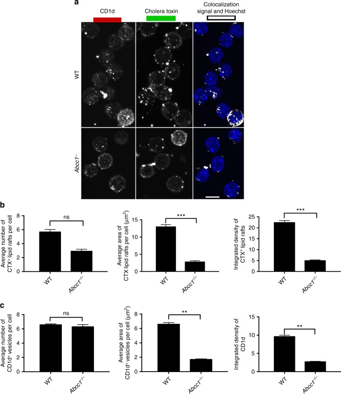
Alterations in CD1d and lipid rafts in Abcc1−/− mice. a Peritoneal macrophages from wild-type (WT) and Abcc1−/− mice were stained with anti-CD1d, Cholera toxin B (CTX) along with Hoechst for staining nucleus. Images were acquired using confocal microscopy. Figure shows representative images for the localization of CD1d in CTX+ compartment in peritoneal macrophages at steady state. b Figure represents the number of Cholera toxin (CTX+) vesicles, average area of CTX+ vesicles, and integrated density of CTX expression. c A compilation of the number of CD1d vesicles, average area of CD1d vesicles, and integrated density of CD1d expression on the cell surface. Representative images from one of two experiments with at least 100 cells per condition. Scale bar 10 μm. Graphs represent mean ± SEM. **p < 0.01; ***p < 0.001 (one-way ANOVA). See also Supplementary Fig. 13

