
Fig. 6.
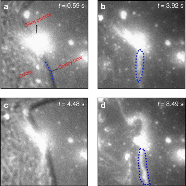
Visualizing MIMS in real-time. Wide-field fluorescence microscopy images of a dye-labelled copolymer 3-coated silica particle at (x = 50 μm, y = 83 μm) in Supplementary Movie 1. a, c Show the overlap of the swarmer colony and labelled copolymer 3 recorded with white and red light irradiation, respectively, and b, d show only the images of copolymer 3. Examples of fluorescent streaks arising from removed copolymer 3 are enclosed within blue dotted lines in (b) and (d). The time of each image t is given. The image contrast is enhanced resulting in particles appearing larger than their actual size due to light scattering