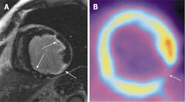
Figure 2.

A sixty-seven-year-old man with severe coronary artery disease and history of myocardial infarction. A: Short axis inversion-recovery gradient-recalled echo cardiac magnetic resonance imaging (CMR) image of the mid- to apical-portion of the left ventricular shows a small area of transmural late gadolinium enhancement (LGE) in the inferolateral wall (broad arrow). CMR viability scores: Anterior: 2; anterolateral: 2; inferolateral: 4, inferior: 2; B: The positron emission tomography (PET) image of the corresponding slice reveals an uptake defect (broad arrow) in the same segment suggesting a transmural scar. PET viability scores: anterior: 2; inferolateral: 4. Small subendocardial scars with LGE in CMR (A) in the anterolateral and inferior wall (small arrows) were overseen in PET.
