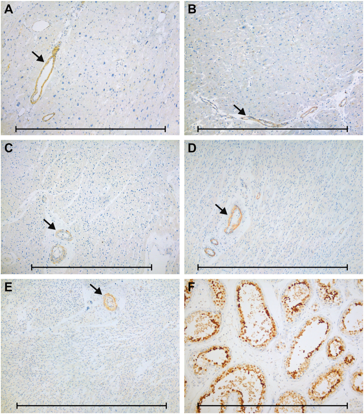
Fig. 2.
Examples of immunohistochemical staining for CALR3 (antibody NBP2-33390) in normal and affected tissues. No expression in myocardial tissue from (a) patient with c.564del-positive DCM, b patient with ischemic cardiomyopathy, and c child, d newborn, and e fetus with structurally normal hearts. Instead, we observed positive staining of arteriolar smooth muscle cells in all samples (arrows). f High expression of CALR3 in testicular germ cells as positive control tissue. Scale bars: 1 mm

