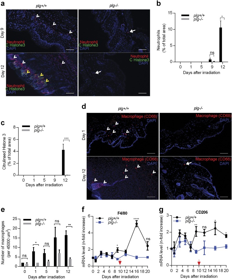
Fig. 3. Large numbers of inflammatory cells infiltrate the irradiated skin of plg+/+ but not plg−/− mice.
a Representative photographs of skin sections from plg+/+ and plg−/− mice on days 9 and 12 after irradiation stained for neutrophils (red), DAPI (blue), and NETs (citrullinated histone H3) (green). White arrowheads and arrows point to neutrophils in plg+/+ and plg−/− mice, respectively. Yellow arrowheads point to NETs in plg+/+ mice. b, c Quantification of neutrophils and NETs in the skin sections from plg+/+ and plg−/− mice on different days after irradiation (n ≥ 3 per genotype). d Representative immunostaining for macrophages (red) in sections from irradiated plg+/+ and plg−/− mouse skin on days 1 and 12 after irradiation. Arrowheads and arrows point to macrophages in plg+/+ and plg−/− mice, respectively. e Quantification of macrophages in the skin sections from plg+/+ and plg−/− mice on different days after irradiation (n ≥ 3 per genotype). Scale bar = 100 µm. f, g Expression of F4/80 and CD206 mRNA, respectively, in skin extracts from plg+/+ and plg−/− mice on different days after irradiation quantified by RT-PCR (n ≥ 4 per genotype). The red arrow indicates the day when dermatitis becomes visible in plg+/+ mice. *P < 0.05; **P < 0.01; ***P < 0.005; ****P < 0.001

