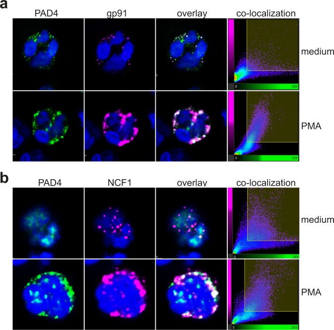
Figure 2.
Co-localization of PAD4 with gp91 and NCF1 in resting or activated neutrophils. (a) Confocal images of resting neutrophils (upper panels) or neutrophils stimulated with 25 nM PMA (lower panels) stained with anti-PAD4 (green) or anti-gp91 (magenta). (b) Confocal images of resting neutrophils (upper panels) or neutrophils stimulated with 25 nM PMA (lower panels) stained with anti-PAD4 (green) or anti-NCF1 (magenta). The fourth panels show the co-location of pixels of the two colors. The data represent the results of three independent experiments.

