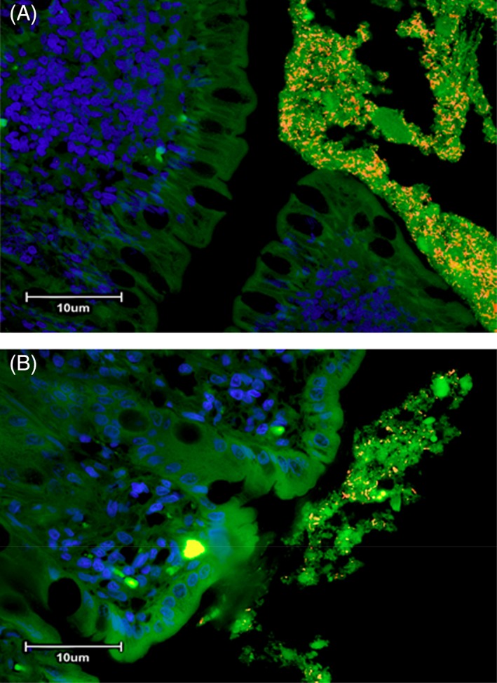
Figure 3.

(A) Three‐color fluorescence in situ hybridization (FISH) of Fusobacterium spp. identified in colonic biopsies of a cat with small cell GI LSA. Cy‐3‐labeled Fusobacterium spp. (labeled orange) localized within adherent mucus of a colonic biopsy specimen. The mucus is also occupied by other bacteria (total bacteria labeled green with FITC‐Eub). The dark blue structures are epithelial cell nuclei stained with DAPI. (B) Three‐color fluorescence in situ hybridization (FISH) of Fusobacterium spp. identified in colonic biopsies of a cat with IBD. Cy‐3‐labeled Fusobacterium spp. (labeled orange) localized within adherent mucus of a colonic biopsy specimen. The mucus is also occupied by other bacteria (total bacteria labeled green with FITC‐Eub). The dark blue structures are epithelial cell nuclei stained with DAPI
