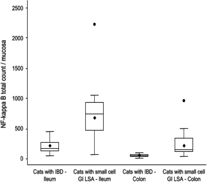
Figure 6.

Mucosal NF‐κB cellular expression in GI biopsies of cats with IBD compared to cats with small cell GI LSA in the ileum and colon (total = 28 cats; IBD = 14 cats; small cell GI LSA = 14 cats). Boxes show lowest, median, and upper quartiles. Whiskers represent 1.5 of the interquartile range, with means denoted by the black rhombi and outliers denoted by the black circles. Median NF‐κB mucosal expression is significantly higher in the ileal (median = 750; P = .0055) and colonic (median = 154; P < .0001) biopsies of cats with small cell GI LSA when compared to the ileal (median = 169) and colonic (median = 49.5) biopsies of cats with IBD
