
Figure 1.
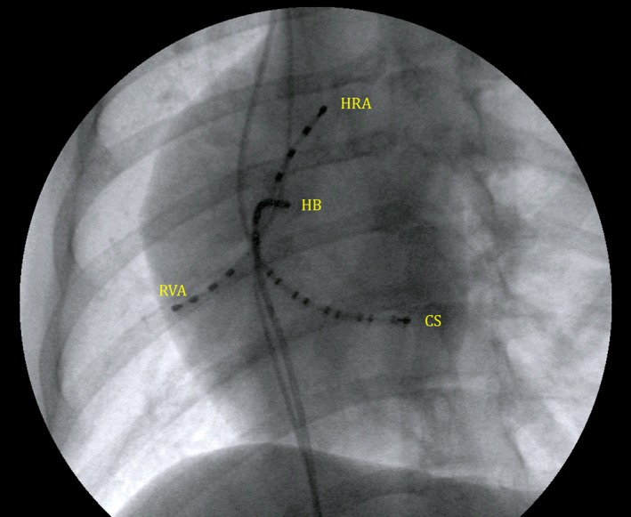
A 30° left anterior oblique fluoroscopic view of multielectrode catheter positioning for the initial EP study. HRA, high right atrium; HB, His bundle region; CS, coronary sinus; RVA, right ventricular apex. All multielectrode catheter poles are labeled by convention in numerical order starting at the distal (tip) electrode