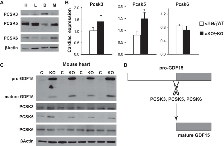
FIG 6.
PCSK3, -5, and -6 expression in mouse models of heart disease. (A) Representative Western blot images of PCSK3, PCSK5, and PCSK6 proteins in the heart (H), liver (L), brown fat (B), and gastrocnemius muscle (M) of 16-day-old control αHet/γWT mice. (B) Expression of Pcsk3, Pcsk5, and Pcsk6 in the hearts of control αHet/γWT and cardiac ERRα/γ KO (αKO/γKO) mice (n = 6 to 8). *, P < 0.05. (C) Representative Western blot images of GDF15, PCSK3, PCSK5, and PCSK6 proteins in the hearts of littermate control αHet/γWT and cardiac ERRα/γ KO mice (n = 4). (D) Diagram summarizing findings of pro-GDF15 cleavage by PCSK3, -5, and -6. β-Actin served as a loading control in panels A and C.

