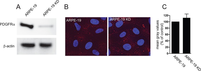
FIG 6.
Binding of soluble trimer to ARPE-19 cells is not affected by silencing PDGFRα. (A) Cell lysates from normal ARPE-19 cells or ARPE-19 KD cells expressing an shRNA targeting PDGFRα were analyzed by Western blotting using anti-PDGFRα polyclonal antibody 3164S (R & D Systems) according to the manufacturer's instructions. The membrane was also probed with a monoclonal antibody to β-actin (Sigma AC-74) to serve as a loading control. (B) Normal ARPE-19 cells or ARPE-19 KD cells were incubated with soluble trimer complexes (100 μg/ml) for 1 h at 4°C. The cells were then washed once in cold PBS and then fixed with 4% paraformaldehyde and processed for immunofluorescence microscopy. Protein complexes were detected using anti-gH MAb 14-4b followed by Dylight-594 goat anti-mouse secondary antibody. The cells were counterstained with DAPI (4′,6-diamidino-2-phenylindole), and the coverslips were mounted onto glass slides with Fluoromount-G. Images were captured by a 60× objective using the Deltavision Core DV Widefield Deconvolution system. (C) Histograms showing the quantification of HCMV soluble trimer binding to cell surfaces of ARPE-19 and ARPE-19 KD cells. The data are displayed as mean gray values (arbitrary units) relative to control conditions as analyzed by ImageJ. Data were collected from three separate images for each condition.

