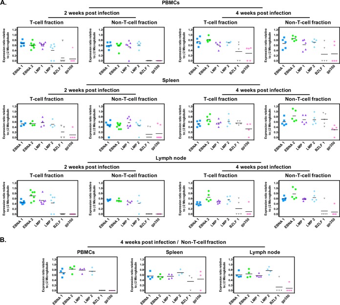
FIG 3.
EBV type 2 gene expression pattern in humanized mouse cell fractions. Purified RNA was isolated from all T cell and non-T cell fractions harvested at 2 and 4 wpi. For all cell fractions that were positive for EBV genome, RNA was reverse transcribed, and biplex PCRs were carried out for EBV genes EBNA-1, EBNA-2, LMP-1, LMP-2, BZLF1, gp350, and human β2-microglobulin. (A) EBV-2-infected mice. Cell fractions were analyzed as follows at 2 wpi: T cell fraction in PBMC, n = 7; non-T cell fraction in PBMC, n = 6; T cell fraction in spleen, n = 4; non-T cell fraction in spleen, n = 7; T cell fraction in LN, n = 7; non-T cell fraction in LN, n = 4. At 4 wpi, n = 5 for all cell fractions. (B) EBV-1-infected mice. At 4 wpi, n = 4 for all non-T cell fractions. The EBV-2 LCL-10 lymphoblastoid cell line was used as a positive control for all latent amplicons, and the EBV-2 Jijoye Burkitt's lymphoma cell line induced to reactivate was used for the lytic amplicon.

