FIG 3.
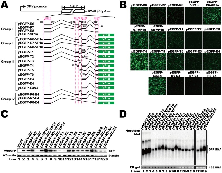
Identification of the cis elements in the 5′ UTR that regulated RNA abundance and translation. (A) Diagram of green fluorescent protein (GFP) reporter plasmids. Group I plasmids contained either the entire 5′ UTR or VP1 unique sequences. Group II plasmids contained both the 5′ UTR and VP1u. Group III constructs harbored VP1u with deletions in exons 1 to 4. Exon 4 in the plasmids of group IV was deleted. SV40, simian virus 40. (B) Microscopy of the fluorescent signal when reporter plasmids were transfected. (C) Western blot. The lysates of transfected HEK293T cells were analyzed using anti-GFP antibody, and β-actin served as the loading control. (D) Northern blot. Total RNAs were harvested from transfected cells, and Northern blotting was carried out as described in the legend to Fig. 2D. Ethidium bromide (EB)-stained 18S RNA bands are shown as the loading control. The image in panel D was spliced from two different gels because the samples could not be run in one gel.

