Fig. 2.
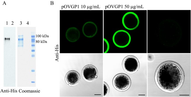
pOVGP1 expressed by mammalian cells binds to ZP of bovine presumptive zygotes. A. pOVGP1 was expressed in HEK 293T cells, purified, separated by SDS-PAGE, and analyzed by immunoblot using a monoclonal antibody against the His tag and Coomassie blue stain. Bands of ~90 kDa correspond to reduced pOVGP1 (lanes 1 and 3) were detected. Lanes 2 and 4 correspond to the negative control. B. Bovine presumptive zygotes incubated in IVF medium containing pOVGP1 at 10 or 50 µg/ml and in IVF medium without pOVGP1 were fixed and imaged by confocal fluorescence microscopy using a monoclonal antibody against the His tag (n = 15). Bright immunofluorescence was detected throughout the entire thickness of the ZP. The intensity was higher in the group supplemented with 50 µg/ml of pOVGP1 than in the 10 µg/ml group. No fluorescent signal was found in the untreated group. Scale bars = 50 µm.

