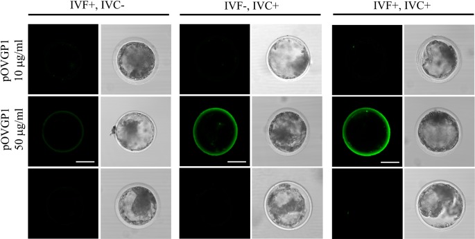
Fig. 3.
pOVGP1 is detected in the ZP of D9 blastocyst when IVC medium is supplemented with 50 µg/ml of pOVGP1. On Day 9, blastocysts were recovered, fixed, and imaged by confocal fluorescence microscopy using a monoclonal antibody against the His tag (n = 21). Bright immunofluorescence was only detected in the ZP of the blastocysts when IVC medium was supplemented with 50 µg/ml of pOVGP1. No fluorescent signal was found in the groups supplemented with 10 µg/ml of pOVGP1, or in the untreated group. Scale bars = 50 µm.

