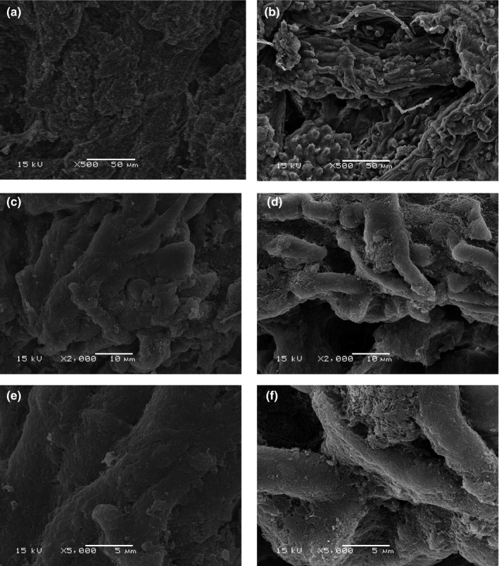
Figure 4.

Scanning electron microscopy (SEM) photographs from control and ultrasonic‐treated whelk meat. (a, c, e) Whelk meat samples without ultrasonic treatment; (b, d, f) whelk meat samples subjected to ultrasonic treatment. The magnification was set as ×500 (a, b), ×2,000 (c, d) and ×5,000 (e, f)
