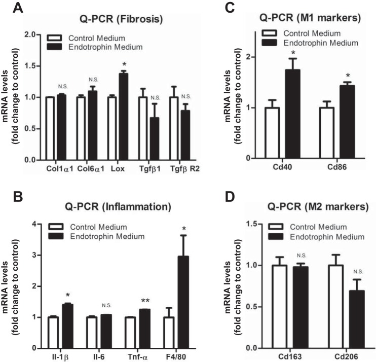
Fig. 5.
Endotrophin upregulated LOX and proinflammatory and classically activated marker (M1) genes in macrophages. A: qPCR analysis of mRNA levels of fibrotic genes Col1α1, Col6α1, Lox, Tgfβ1, and TgfβR2 in macrophages treated with control or endotrophin-conditioned medium (data are means ± SE; n = 3. Student’s t-test, *P < 0.05). B: qPCR analysis of mRNA levels of inflammatory genes IL-1β, IL-6, TNFα, and F4/80 in macrophages treated with control or endotrophin-conditioned medium (data are means ± SE; n = 3. Student’s t-test, *P < 0.05, **P < 0.01). C: qPCR analysis of mRNA levels of M1 macrophage markers Cd40 and Cd86 in macrophages treated with control or endotrophin-conditioned medium (data are means ± SE; n = 3. Student’s t-test, *P < 0.05). D: qPCR analysis of mRNA levels of M2 macrophage markers Cd163 and Cd206 in macrophages treated with control or endotrophin-conditioned medium (data are means ± SE; n = 3. Student’s t-test).

