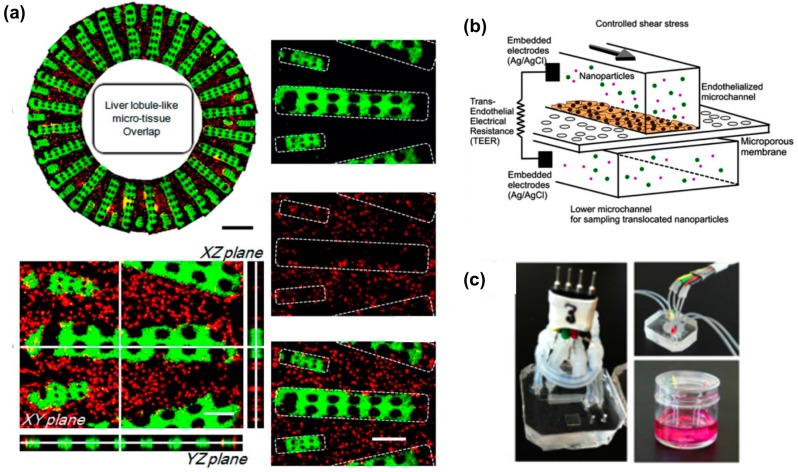
Figure 1.
(a) Geometrical 3D arrangement of a liver lobule-on-a-chip. An hydrogel patterning technique is employed by Ma et al. to obtain radially distributed hepatocytes (green) in a network of endothelial cells (red). Adapted from [42] with permission; (b) 3D sketch of a blood–brain barrier microdevice model with upper compartment hosting endothelial monolayer culture on a microporous membrane and lower compartment for collection of transported nanoparticles. Electrical measurements across the endothelial monolayer provide information on barrier integrity; (c) Pictures of blood–brain barrier microdevices with fluidic and electical connections (adapted from [52] with permission).

