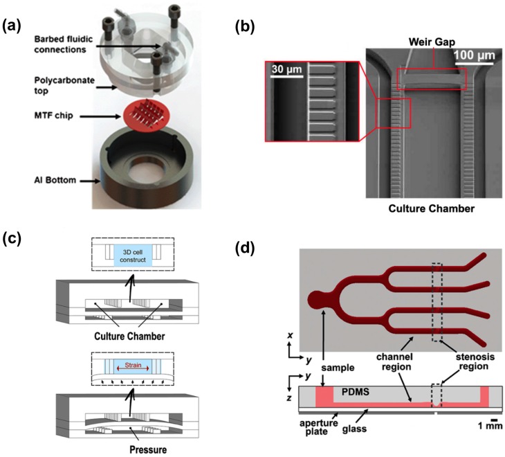
Figure 2.
Cardiovascular organs-on-a-chip. (a) Exploded view of heart-on-a-chip device. Adapted from [96] with permission from The Royal Society of Chemistry; (b) Scanning electron micrograph of the microphysiological system. Red rectangular boxes show the 2 mm endothelial-like barrier and the weir gap. Adapted with permission from [103]; (c) Schematic of the 3D beating heart-on-a-chip microdevice. The polydimethylsiloxane (PDMS) membrane between compartments deforms, compressing the 3D cell construct. Adapted from [87] with permission from The Royal Society of Chemistry; (d) Schematic showing top and side view of the microfluidic device for inducing platelet aggregation at four distinct high shear stenotic regions. Adapted with permission from [104].

