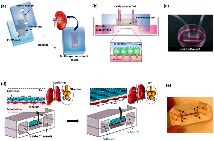
Figure 3.
Biological barrier microfluidic models applied for kidney and lung organs. Kidney platform: (a) sketch of the fabrication process involving the sandwich assembly of the porous membrane between two PDMS layers; (b) schematic of the chip operating principle; and (c) picture of the device connected to a syringe pump through silicon tubing. Adapted from [137] with permission. Human lung on a chip: (d) schematic of the epithelial and endothelial cells co-cultured on opposite sides of the porous membrane, stretched applying vacuum in the side channels to mimic alveolar deformation during normal breathing; and (e) picture of a microdevice filled with color dyes. Adapted from [121] with permission.

