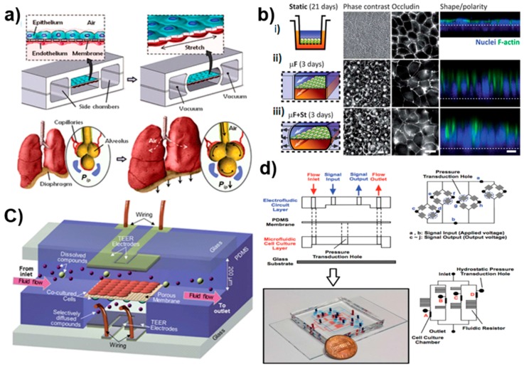
Figure 3.
Examples of sophisticated organ-on-chip applications. (a) Compartmentalized polydimethylsiloxane (PDMS) microchannels for breathing activities in the lung. A thin, porous, and flexible PDMS membrane coated with the extracellular matrix (ECM) forms an alveolar-capillary barrier. The device recreates physiological breathing movements by applying a vacuum to side chambers and leads to mechanical stretching of the PDMS membrane to form the alveolar-capillary barrier. The inhalation in the living lung contracts the diaphragm and reduces the intrapleural pressure and physical stretching of the alveolar-capillary interface [78]; (b) The morphology of epithelial cells cultured in the (i) static Transwell system for 21 days, gut-on-a-chip with a microfluidic flow without (ii) or with (iii) the application of cyclic mechanical deformation for three days. The schematic layout of (left); fluorescence views (center) of the occludin as the tight junction (TJ) protein, and the confocal fluorescence views (right) of the epithelium (nuclei in blue and F-actin in green) [80]; (c) The design of the developed microfluidic blood-brain barrier (BBB) with integrated electrodes for measuring the trans-epithelial resistance across the barrier [98]; (d) The microfluidic cell culture device with embedded electrofluidic pressure sensors. The PDMS membrane is sandwiched between two other PDMS layers: an electrofluidic circuit layer and a microfluidic cell culture layer. The layout of the electrofluidic circuit layer for pressure sensing at four locations, and an equivalent Wheatstone bridge circuit of the pressure sensor [84].

