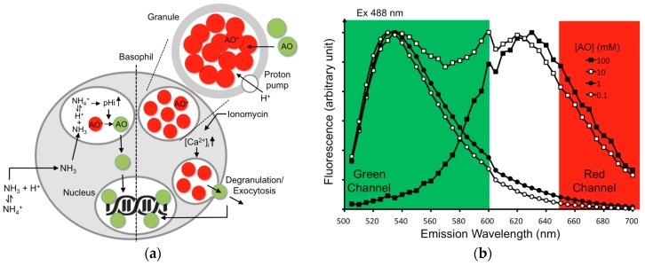
Figure 1.
AO are trapped in the granules of basophils and exhibit red shift at high concentrations. Weakly basic AO molecules are selectively accumulated within granules as a result of the intragranular acidic pH. Upon entering the basophilic granules, AO dyes become protonated and are trapped inside the organelles (a, right). The resulting high concentrations of AO cause a red shift, thereby giving red fluorescence after excitation at 488 nm. When challenged with NH4Cl, NH4Cl decomposes into ammonia in solution that passes through membranes easily, accumulating in the basophilic granules and promoting granular alkalinization to release AO (a, left). When challenged with ionomycin, the intracellular Ca2+ ion level ([Ca2+]i) increases, which triggers degranulation and releases AO externally (a, right). The released AO dyes then bind to the nuclear DNA and generate strong green fluorescence (a). To demonstrate the phenomenon of red shift, AO was dissolved at the concentration (mM) as indicated (0.1 (open circle), 1 (solid circle), 10 (open square), 100 (solid square)). Emission spectrum of AO at each concentration was scanned from 500 to 700 nm with excitation at 488 nm; emission peak from the solutions of various AO concentrations was normalized to the same level for comparison because the quantum efficiency of the AO red fluorescence is much weaker than that of the green one. High concentrations of AO (≥10 mM) show a red shift (b).

