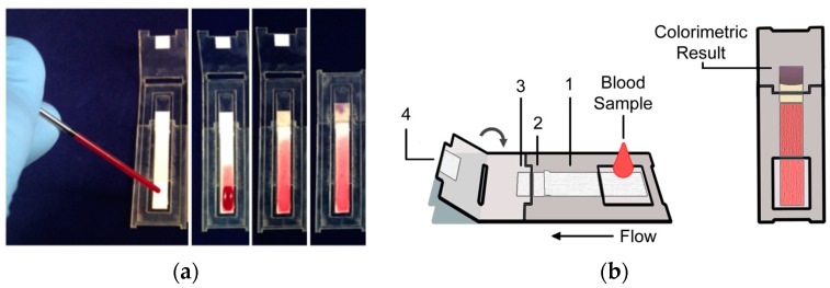
Figure 1.
Prototype disposable paper-based device. (a) Image series showing different stages of a device run. From left to right, 40 μL of whole blood is applied to the device using a disposable glass capillary and plunger (PTS Diagnostics; left), blood flows into the plasma separation membrane, plasma is collected in the pads immediately downstream and the enzymatic reaction takes place, and after the device is folded closed, the colorimetric reaction takes place and signal develops; (b) Schematic of the disposable device. The substrate labeled 1 is the blood to plasma separation membrane, substrates 2 and 3 house the enzymatic reaction, and substrate 4 houses the colorimetric reaction. (Right) The device is folded at ~6 min and then read at ~8 min using an instrument for a semi-quantitative result.

