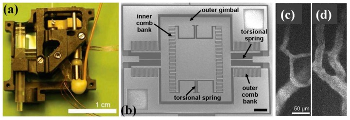
Figure 16.
MEMS based two-photon microscope. (a) Photograph of the assembled microscope, electrical lines control the MEMS scanner and focusing micromotor. (b) Scanning electron micrograph of the two-dimensional MEMS scanner for en-face two-photon imaging, 750 µm × 750 µm scanning mirror in a 3.2 mm × 3.0 mm die, six banks of vertical comb actuators drive the mirror, which has a gimbal design, scale bars are 250 µm. (c,d) Images of neocortical capillaries, averaged over eight frames acquired over 2 s at 4 Hz (Reproduced with permission from OSA [99]).

