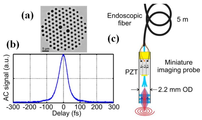
Figure 17.
Scanning fiber based two photon endomicroscope system. (a) Close view of the inner core of the custom-design air-silica DC-PCF through scanning electron microscopy (SEM). Pure silica is in grey and air in black. (b) Second order autocorrelation (AC) of the IR excitation pulse at the exit of the 5-m-long endoscopic fiber for a delivered power of 20 mW. The pulse duration has been calculated from the AC duration by using the suitable conversion factor (i.e., 1.54 = (AC duration)/(pulse duration) at FWHM. Accordingly, the pulse duration was equal to 39 fs (FWHM). (c) Scheme of the miniature fiber scanning imaging probe which is embedded inside a 2.2 mm outer diameter (OD) stainless steel biocompatible tube (Reproduced with permission from Nature Publisher Group [107]).

