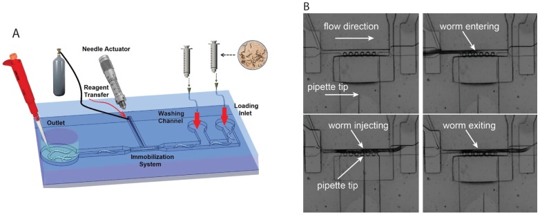
Figure 4.
Microfluidic devices for microinjection in (A) closed microchannels (Ghaemi [41] and (B) open chambers (Song et al. [43]. Panel (B) reproduced with permission from American Institute of Physics Publishing. The device in panel (A) contains worm loading and washing channels (on the right) and an outlet for collecting injected worms. Worm is immobilized in the middle region for injection. The image frames in panel (B) show a sequence of worm loading, injection, and flushing. Refer to respective references for more details.

