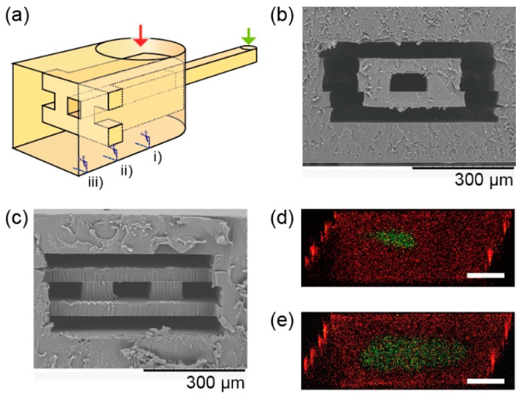
Figure 6.
Cross-sectional images of a 3D sheath device. (a) Conceptual image of the 3D sheath device. (b) A SEM image of a cross section of the device at position (i) in Figure 6a, and (c) at position (ii) in Figure 6c. (d) A confocal microscope image of the 3D sheath at position (iii) with the flow rate of fluorescein as 90 µL/min and rhodamine as 450 µL/min. (e) Fluorescein as 40 µL/min and rhodamine as 210 µL/min. Scale bars are 100 µm.

