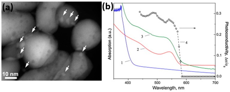
Figure 55.
(a) High angular annular dark field scanning transmission electron microscopy (STEM) image of CdSe QD1/ZnO sample. CdSe nanoparticles are marked with arrows; (b) Absorption spectra of ZnO powder (1); CdSe QDs suspended in solution (2); QD3/ZnO powder (3); and dependence of photoconductivity of QD3/ZnO sample on the excitation wavelength (4). Reproduced with permission from [78].

