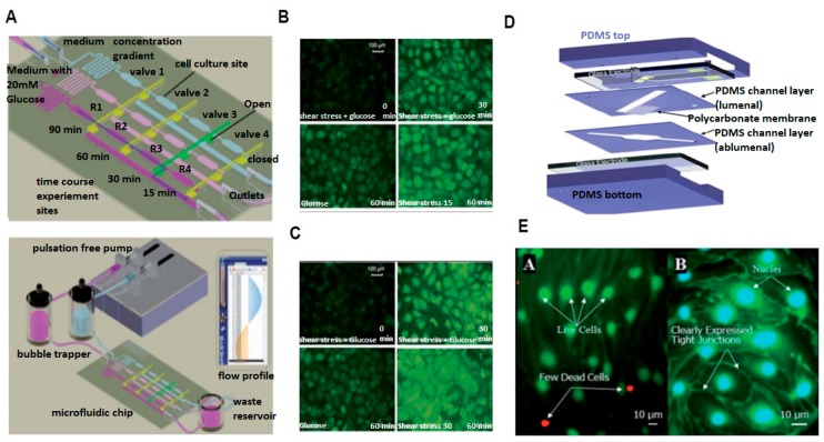
Figure 2.
2D Microdevices Utilized to Study Glucose Exposure and the Blood Brain Barrier Under Fluid Flow. (A) Schematic of microfluidic chip for exposing cells to various glucose concentrations under fluid flow; (B) Fluorescent image of endothelial cell morphology and reactive oxygen species spatial distribution under low fluid shear stress; (C) Fluorescent image of endothelial cell morphology and reactive oxygen species spatial distribution under high fluid shear stress; (D) Schematic of μ-Blood Brain Barrier device showing; and (E) fluorescent images of cells cultured inside μ-Blood Brain Barrier device (adapted from Chin et al. [16] and Booth et al. [17]).

